Brain structure and function
Brain structure and function were assessed in 2018–2019 among 45 adolescents aged 18-19 years participating in the FitBrain study, a PANIC substudy.
Brain structure was examined using three-dimensional T1-weighted magnetic resonance imaging (MRI) with a 3-Tesla scanner (Philips Achieva). Structural MRI data were used to quantify regional gray matter volumes, including cerebellar gray matter and its lobules, as well as cortical thickness.
White matter microstructure was characterized using diffusion tensor imaging (DTI), including measures of fractional anisotropy, mean diffusivity, axial diffusivity, and radial diffusivity.
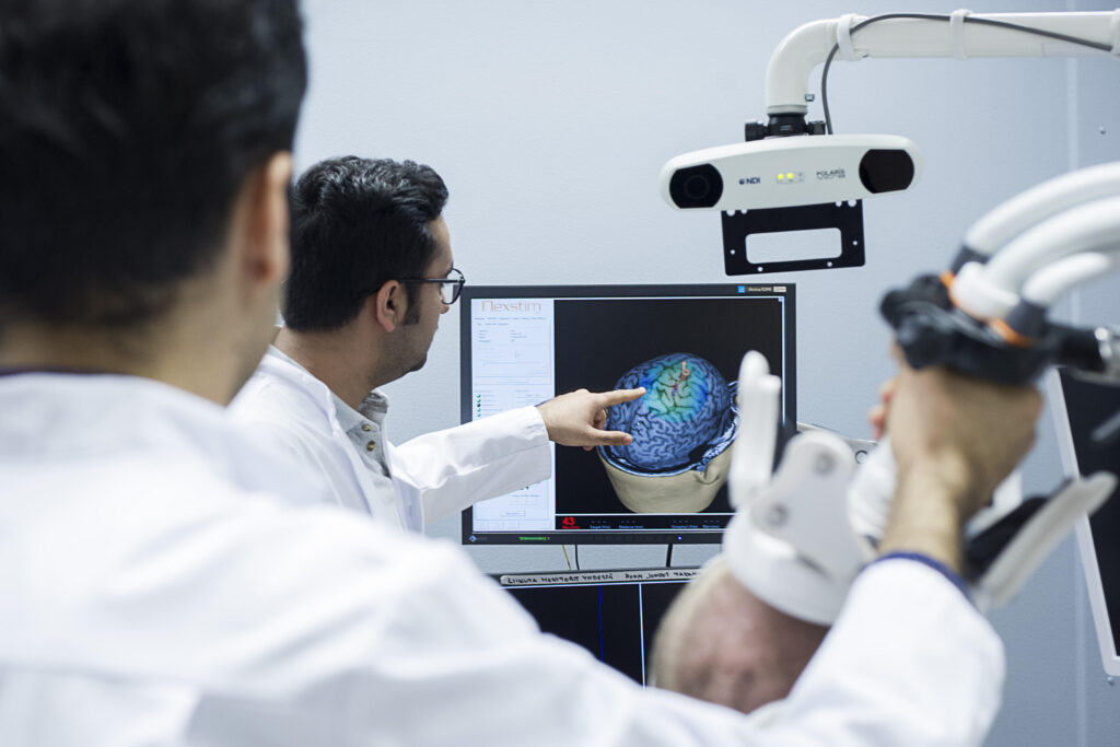
Motor cortical function was assessed using navigated transcranial magnetic stimulation (nTMS) to characterize cortical excitability and inhibition. Measurements included resting motor threshold, motor-evoked potential amplitude, cortical silent period, silent period threshold, and long-interval intracortical inhibition.
Together, these complementary methods provided detailed information on brain morphology, white matter microstructure, and cortical neurophysiological function during adolescence.