Adipose tissue imaging
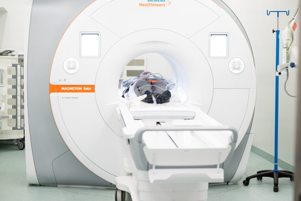
Abdominal visceral and subcutaneous adipose tissue were assessed using magnetic resonance imaging (MRI) at the 16-year follow-up, when participants were young adults aged 23–25 years.
Magnetic resonance imaging (MRI) enables non-invasive quantification of abdominal adipose tissue depots and provides detailed information on body fat distribution. These measurements are complemented by assessments of whole-body composition, glucose and lipid metabolism, and molecular characteristics of subcutaneous adipose tissue.
Together, these data enable investigation of how adipose tissue distribution and molecular function are associated with systemic metabolic health, insulin resistance, other disturbances in glucose and lipid metabolism, and liver fat accumulation in early adulthood.